 If you have a symptom that suggests lung cancer, your doctor must find out whether it’s from cancer or something else. Your doctor may ask about your personal and family medical history. Your doctor may order blood tests, and you may have one or more of the following tests:
Physical exam: Your doctor checks for general signs of health, listens to your breathing, and checks for fluid in the lungs. Your doctor may feel for swollen lymph nodes and a swollen liver.
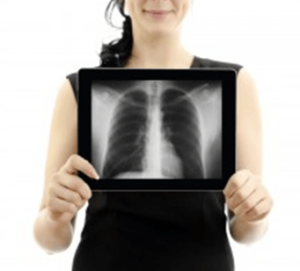
Chest x-ray: X-ray pictures of your chest may show tumors or abnormal fluid.
CT scan: Doctors often use CT scans to take pictures of tissue inside the chest. An x-ray machine linked to a computer takes several pictures. For a spiral CT scan, the CT scanner rotates around you as you lie on a table. The table passes through the center of the scanner. The pictures may show a tumor, abnormal fluid, or swollen lymph nodes.
The only sure way to know if lung cancer is present is for a pathologist to check samples of cells or tissue. The pathologist studies the sample under a microscope and performs other tests. There are many ways to collect samples. Currently, the United States Preventive Services Task Force (USPSTF) recommends annual screening for lung cancer with low-dose computed tomography (LDCT) in adults aged 55 to 80 years who have a 30 pack-year smoking history and currently smoke or have quit within the past 15 years. Screening should be discontinued once a person has not smoked for 15 years or develops a health problem that substantially limits life expectancy or the ability or willingness to have curative lung surgery. (Click here to learn more about lung cancer screening.)
Your doctor may also order one or more of the following tests to collect samples:
Sputum cytology: Thick fluid (sputum) is coughed up from the lungs. The lab checks samples of sputum for cancer cells.
Thoracentesis: The doctor uses a long needle to remove fluid (pleural fluid) from the chest. The lab checks the fluid for cancer cells.
Bronchoscopy: The doctor inserts a thin, lighted tube (a bronchoscope) through the nose or mouth into the lung. This allows an exam of the lungs and the air passages that lead to them. The doctor may take a sample of cells with a needle, brush, or other tool. The doctor also may wash the area with water to collect cells in the water.
Fine-needle aspiration: The doctor uses a thin needle to remove tissue or fluid from the lung or lymph node. Sometimes the doctor uses a CT scan or other imaging method to guide the needle to a lung tumor or lymph node.
Thoracoscopy: The surgeon makes several small incisions in your chest and back. The surgeon looks at the lungs and nearby tissues with a thin, lighted tube. If an abnormal area is seen, a biopsy to check for cancer cells may be needed.
Thoracotomy: The surgeon opens the chest with a long incision. Lymph nodes and other tissue may be removed.
Mediastinoscopy: The surgeon makes an incision at the top of the breastbone. A thin, lighted tube is used to see inside the chest. The surgeon may take tissue and lymph node samples.
CT scan: CT scans may show cancer that has spread to your liver, adrenal glands, brain, or other organs. You may receive contrast material by mouth and by injection into your arm or hand. The contrast material helps these tissues show up more clearly. If a tumor shows up on the CT scan, your doctor may order a biopsy to look for lung cancer cells.
Bone scan: A bone scan may show cancer that has spread to your bones. You receive an injection of a small amount of a radioactive substance. It travels through your blood and collects in your bones. A machine called a scanner detects and measures the radiation. The scanner makes pictures of your bones on a computer screen or on film.
MRI: Your doctor may order MRI pictures of your brain, bones, or other tissues. MRI uses a powerful magnet linked to a computer. It makes detailed pictures of tissue on a computer screen or film.
PET scan: Your doctor uses a PET scan to find cancer that has spread. You receive an injection of a small amount of radioactive sugar. A machine makes computerized pictures of the sugar being used by cells in the body. Cancer cells use sugar faster than normal cells and areas with cancer look brighter on the pictures.